Introduction
A perineal hernia occurs when tissue or organs protrude through a defect of the pelvic floor. They can be congenital or acquired after surgery or trauma in the pelvic area .1,2 Abdominoperineal resection of the rectum and pelvic exenteration are the most common causes of postoperative perineal hernias with an incidence of secondary perineal hernia between 1 to 13%.2 Symptoms of a perineal hernia often include perineal discomfort, pain, urinary dysfunction, and rarely, bowel obstruction.2 Treatment of perineal hernias depends on the patient’s level of bother. In patient’s that are not bothered by their symptoms, treatment is usually conservative. However, some patients may elect for surgical treatment due to the significant impact on quality of life. Surgical repair of perineal hernias is complex due to the challenging anatomic location of the defect and often requires a multidisciplinary approach.2 Due to the relative rarity, there is no consensus regarding the optimal treatment.2
Perineal hernias containing gynecological organs such as the uterus and cervix are even more rare, and only a few such case reports have been previously described. A prior case study with an extremely large, 15 x 9cm, perineal uterine-containing hernia involved repair with total hysterectomy, right-sided adnexectomy, vaginal vault suspension, and colpoperineorrhaphy.3 However, this patient’s surgery was completed via laparotomy with an infraumbilical approach and had a recurrence after 2 months.3
Here, we demonstrate a multidisciplinary approach to surgically correct a complex perineal hernia and pelvic organ prolapse. To our knowledge, surgical treatment involving a mesh-augmented perineal hernia repair with uterine-preserving, extraperitoneal hysteropexy has not been described previously.
Case Summary
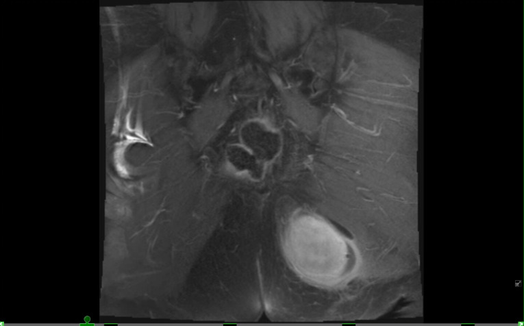
The patient is a 63-year-old G3P2012 with a history of a perirectal abscess and fistula. She underwent an exploratory laparotomy, lysis of adhesions, takedown of high rectal fistula, and flexible sigmoidoscopy in December 2024. One month later, she presented to her primary care provider with abdominal pain and reports of a bothersome left gluteal mass. Her provider ordered a pelvic MRI which showed a large left perineal hernia containing the uterus and cervix with a defect involving the left pelvic floor musculature (Figure 1).
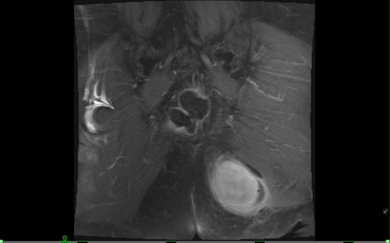
The patient was referred to urogynecology for evaluation of the pelvic floor hernia. Physical examination demonstrated a palpable, reducible mass in the left gluteal region. On vaginal exam, the uterus and cervix were herniated into the left perirectal fossa, 3-4 cm proximal to the hymen. With gentle manipulation, the cervix and uterus could be manipulated into the vaginal canal by applying pressure to the left gluteal mass. She was noted to have stage 2 uterovaginal prolapse.
The patient was then referred to a specialized general surgeon with an expertise in abdominal core and hernia reconstruction. After discussion with both urogynecology and general surgery, the patient was recommended a mesh-augmented pelvic floor reconstruction along with uterovaginal prolapse repair. Given the patient’s extensive abdominal surgical history and benign gynecologic histories, she was recommended an extraperitoneal approach with right sacrospinous ligament hysteropexy and posterior colporrhaphy with perineorrhaphy. Preoperative urodynamic testing demonstrated occult stress urinary incontinence. She elected to undergo concomitant urethral bulking at the time of the prolapse repair due to her active tobacco use.
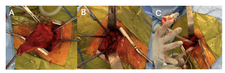
In December 2025, the patient underwent surgical repair of the 8 cm (craniocaudal) x 4 cm (transverse) perineal hernia. First, the patient was placed in right lateral decubitus position, and general surgery made a vertical incision to the left of midline well away from the anus over the hernia sac. The sac was dissected free from the subcutaneous attachments circumferentially. An 8 cm PhasixTM ST mesh was placed on the visceral side and fixed to the pelvic floor musculature using two 0-PDS sutures and adhesive fixation. The pelvic floor musculature was closed using #2 DuraMeshTM suture (Figure 2). Duramesh was used given its increased surface area compared to regular suture, which is particularly advantageous when suturing muscle to muscle. After concluding the hernia repair, the patient was repositioned in dorsal lithotomy, and the sacrospinous ligament hysteropexy, posterior repair, perineorrhaphy, cystoscopy, and urethral bulking were performed. The uterus and cervix were displaced to the level of the right ischial spine using three sutures of 0-PDS. The posterior colporrhaphy and perineorrhaphy were performed in the usual fashion. The operation was uncomplicated, and complete resolution of her vaginal prolapse was achieved. Her immediate postoperative course was unremarkable, and she was discharged on post-operative day 1.
_dissection_of_.png)
Her postoperative course remained uncomplicated. She presented to the urogynecology office at 6 weeks postoperatively for follow-up. She reported extreme satisfaction with resolution of her incontinence and prolapse symptoms since surgery. On physical exam, she continued to demonstrate excellent anterior, apical, and posterior vaginal wall support. CT evaluation 3 months after her repair showed no recurrence and the patient currently remains symptom-free with a significantly improved quality of life.
Discussion
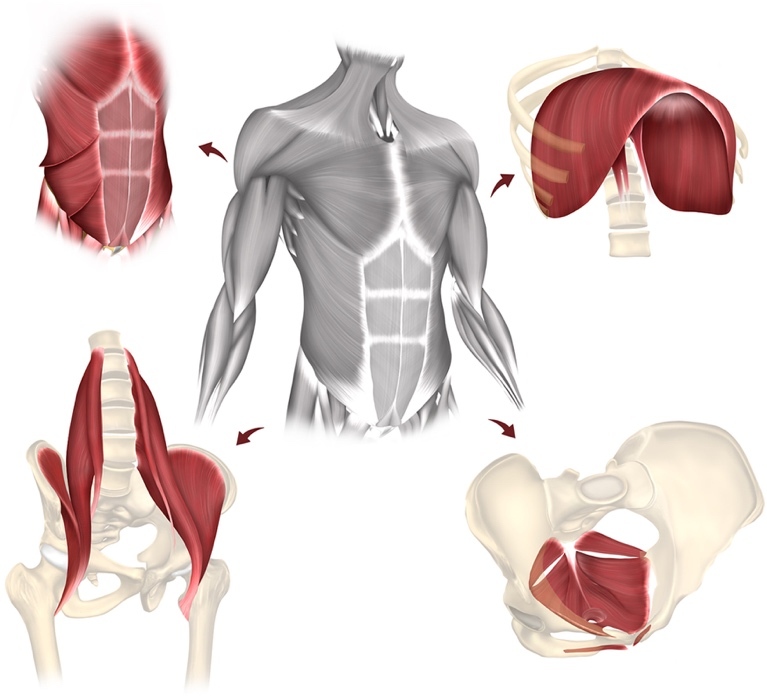
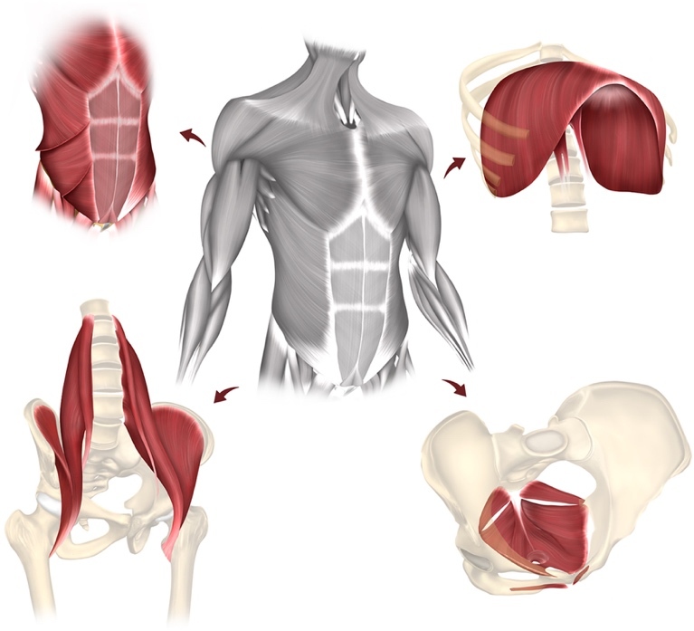
The abdominal core and hernia health are not concepts frequently discussed within the field of urogynecology. However, there is considerable overlap. The term “abdominal core,” encompasses the diaphragm, pelvic floor, and abdominal wall/flanks (Figure 3). Abdominal core health describes the stability and function of the abdominal core and its relation to quality of life.4 Important factors to maintain the abdominal core include prevention of disease by maintaining core strength and a healthy weight, and treatment modalities such as physical therapy and surgery. Further evidence of the interconnection between the abdominal core and urogynecology is the fact that patients who have ventral hernias successfully treated also note improvement in pelvic floor function.4
Hernias can be acquired or congenital. Acquired hernias are categorized as primary or secondary.2 Primary perineal hernias occur in the absence of prior perineal surgery, conditions that increase intra-abdominal pressure, such as pregnancy, or chronic local infections. These are incredibly rare. Secondary perineal hernias are more common and typically arise following perineal surgery or infection.2 Repair of secondary perineal hernias has traditionally focused on closure of the pelvic floor defect using primary closure, biological and synthetic mesh closure, or tissue flap reconstruction through an abdominal, perineal, or combined approach.3,5 However, recurrence rates after perineal hernia repair are high.3,5 A meta-analysis evaluated recurrence rates after biological versus synthetic mesh repair, showing superior outcomes with synthetic mesh (29% recurrence) compared to biological mesh (39%). Despite these high recurrence rates, use of synthetic or biologic mesh reduces recurrence compared to primary repair without mesh.2
Uterovaginal prolapse is a herniation of the pelvic organs due to loss of pelvic floor support. Surgical treatment options for prolapse include a native-tissue or mesh-augmented repair.6,7 If apical prolapse is present, a hysterectomy may or may not be performed depending on examination findings, perioperative comorbidities, gynecologic and family history, surgical considerations and patient autonomy.8,9 Similar to hernia literature, a mesh-augmented approach has a lower recurrence and reoperation rate.6 However, the use of mesh carries its own specific risks, particularly in smokers, where there is a significantly higher rate of mesh complications.7 For our specific patient, a mesh-augmented prolapse repair was not recommended given both her extensive smoking and surgical histories.
Uterine-sparing surgery to treat prolapse is as equally effective and durable as hysterectomy-based cuff suspension procedures.8–11 Hysteropexy procedures also have shorter operative times and decreased blood loss, compared to native tissue repairs with concomitant hysterectomy.9–11 Sacrospinous ligament hysteropexy is an increasingly popular uterine-sparing surgical method for treatment of prolapse and has the added benefit of an extraperitoneal approach in patients with complicated abdominal surgical histories.9–11 Due to our patient’s many medical comorbidities and surgical history, an extraperitoneal approach was recommended to decrease the operative time and reduce the risk of surgical injury. The decision to repair the uterovaginal prolapse at the same time as the perineal hernia repair was two-fold. First, we avoided the perioperative risks of a secondary surgery. Secondly, correcting the hernia defect and uterovaginal prolapse at the same time limited the tension and pressure from the uterus on the healing suture-lines of the hernia repair.
This case highlights the importance of recognizing abdominal core health and its overlap within the field of urogynecology. For our patient, the use of sacrospinous ligament hysteropexy together with a mesh-augmented perineal hernia repair is a novel surgical approach. To our knowledge, this is the first case report detailing a complex multi-disciplinary approach to managing uterovaginal prolapse utilizing a uterine-sparing technique concurrently with a pelvic floor hernia repair and reconstruction. Given the rarity of uterine herniation through postoperative pelvic floor defects, we hope our case will provide useful techniques and considerations for future surgeons treating this complex condition.