Introduction
Kaposiform lymphangiomatosis (KL) is a very rare generalized lymphatic anomaly that presents most commonly in children.1–3 Tumors occur most frequently in the chest with pulmonary or bleeding symptoms,2 although it has been documented in adult patients and can also be found in the retroperitoneum and elsewhere, although rarely without mediastinal involvement.1,4,5 It was first described at the Pediatric Pathology Society meeting in 2004, with the first publications in 2013.2,4,6 KL is defined by abnormal lymphatic channels with “kaposiform” clusters of spindled lymphatic endothelial cells.6 The clinical course can be aggressive and morbid, and treatment usually involves a medical and surgical approach without standardization due to the varied anatomic locations, patient age, and rarity.
Case Summary
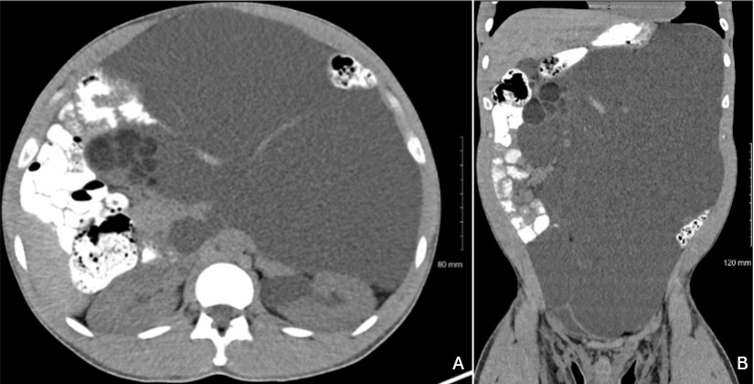
We report a case of a 32-year-old man who was found to have a large cystic abdominal mass after presenting with months of worsening abdominal distension, occasional shortness of breath, and chest discomfort over the course of several months. He was offered open resection for definitive diagnosis and treatment. The patient authorized disclosure of information for this report.
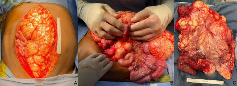
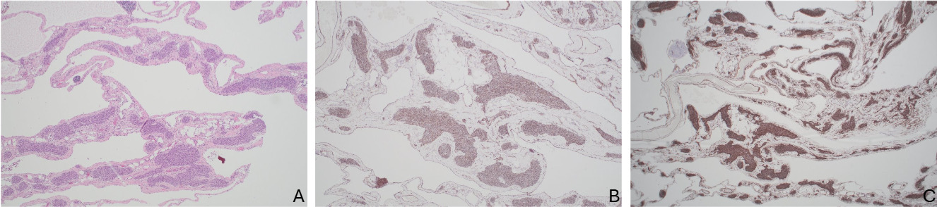
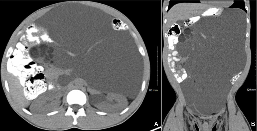
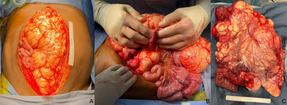
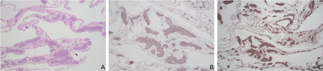
Representative images from pre-operative imaging and operation are shown for reference (Figures 1 and 2). The patient underwent exploratory laparotomy with small bowel resection en block with the tumor. Intraoperatively the mass appeared to originate from within the small bowel mesentery near the fourth portion of the duodenum and had expanded within mesenteric planes from the ligament of Treitz along the transverse colon and descending colon mesentery. The mass was filled almost entirely with lymphatic fluid. The mass was resected with the fourth portion of the duodenum and proximal jejunum while preserving the SMA and proximal jejunal branches. Continuity was restored with a stapled retrocolic duodenojejunostomy between the lateral aspect of D2 and the jejunum, and an appendectomy was performed. Operative time was four hours and 16 minutes. The patient recovered well without complications and was discharged home on post-operative day six. Pathology showed kaposiform lymphangiomatosis. Stains were positive for CD31, CD34, and D2-40. The appendix was normal (Figure 3). Before discharge a chest CT scan was obtained which was normal, showing no intrathoracic pathology.
Discussion
Kaposiform lymphangiomatosis is an extremely rare generalized lymphatic anomaly in adult patients. Presenting symptoms are often secondary to lesion location, such as hemoptysis or pleural effusions for chest lesions, bone or joint pain, or bruising and bleeding from consumptive coagulopathy.6 Abdominal distension as an isolated presenting symptom in an adult patient has not been reported to our knowledge.
Diagnosis of KL is made with a combination of imaging and histopathologic findings. Obtaining tissue can be difficult due to coagulopathy with risk of bleeding, lymphatic leak, or risk of injury to proximal critical structures. Surgery can be both diagnostic and therapeutic.
Histologically KL is characterized by abnormal lymphatic channels containing mural collections of spindled endothelial cells resembling Kaposi sarcoma that stain positively for endothelial markers, including D2-40, PROX1, CD34 and CD31.1,6–8 Most KL have an activating somatic NRAS p.Q61R mutation.7,9 This activating mutation is also seen in human cancers including melanoma, and similar activating RAS mutations are also common in several benign cutaneous lesions (epidermal and congenital melanocytic nevi) and arteriovenous malformations.9 The NRAS mutation activates both the RAS/MAPK and PIK3/AKT/mTOR pathways. Sirolimus, an mTOR inhibitor, has emerged as a potential medical treatment for these tumors.5,8,10
Angiopoeitin-2 is a biomarker that is elevated in KL patients and can be used to differentiate KL from Kaposiform hemangioendothelioma.1,6
Overall prognosis for KL has improved over time. When initially described in 2013, 5-year survival was 51% and overall survival only 34%.4 However recent series report overall survival as high as 79% in one cohort, though this only had 3 patients (n=43) older than 21 years.1
Conclusion
While KL is most commonly found in pediatric patients in the thoracic cavity, KL should be considered in the differential of abdominal cystic masses in adults. Further reporting of this diagnosis is needed to better characterize and treat patients with this incredibly rare anomaly. Surgical resection should be pursued when feasible for both diagnostic and potentially therapeutic purposes.