Introduction
Burn injuries continue to represent a major global public health burden, accounting for significant morbidity, mortality, prolonged hospitalization, disability, psychological trauma, and socioeconomic loss, particularly in low- and middle-income countries.1,2 According to the World Health Organization, the majority of severe burn-related deaths occur in resource-limited settings where access to specialized burn centers, reconstructive services, intensive care support, and rehabilitation remains inadequate.2,3 Young adults are disproportionately affected because of occupational and environmental exposure, especially among rural populations involved in farming, animal herding, open-fire cooking, and outdoor camping activities.3
Flame burns are among the most devastating forms of thermal injury because they frequently involve prolonged exposure and deeper tissue destruction.4 While superficial and partial-thickness burns may heal with conservative management, fourth-degree burns represent the most severe end of the burn spectrum and are characterized by full-thickness destruction extending beyond the skin into subcutaneous tissue, fascia, muscle, tendon, and bone.1,4 Such injuries are uncommon but are associated with extremely high rates of infection, osteomyelitis, sepsis, limb dysfunction, and amputation.5
The lower extremities are particularly vulnerable to complications following deep burn injury because of relatively limited soft tissue coverage over bony prominences such as the tibia.6,7 Exposure of cortical bone presents a major reconstructive challenge, as avascular bone surfaces do not readily support spontaneous granulation tissue formation or skin graft take.7 Delayed wound coverage may further worsen desiccation, necrosis, bacterial colonization, and chronic osteomyelitis.7,8
Management of fourth-degree lower extremity burns requires a multidisciplinary approach involving aggressive resuscitation, infection control, nutritional optimization, serial debridement, metabolic support, soft tissue reconstruction, rehabilitation, and long-term follow-up.1,9 Severe burns also induce a profound hypermetabolic and hypercatabolic response characterized by persistent tachycardia, increased resting energy expenditure, protein breakdown, muscle wasting, immune dysfunction, and delayed wound healing.10,11 Early nutritional rehabilitation and pharmacologic modulation of hypermetabolism, including the use of beta-blockers such as propranolol, have therefore become important adjuncts in modern burn care.10–12
Reconstruction of exposed tibial bone following burn injury remains technically demanding. Various reconstructive strategies have been described including cortical drilling, negative pressure wound therapy, local rotational flaps, regional muscle flaps, free tissue transfer, and split-thickness skin grafting.6–8,13–17 Cortical bone drilling is a useful technique that promotes vascular granulation tissue formation by allowing blood flow from the medullary cavity to reach the exposed cortical surface.7 Once a healthy wound bed is achieved, muscle flaps such as the gastrocnemius and soleus flaps provide durable vascularized coverage and improve resistance to infection while facilitating subsequent skin grafting.13–16
The hemisoleus muscle flap is especially valuable for coverage of middle-third tibial defects because of its reliable segmental vascular anatomy, ease of mobilization, and preservation of partial muscle function.13–16 Similarly, the medial gastrocnemius flap remains one of the most dependable reconstructive options for proximal and upper-middle tibial exposure.8,14 In extensive bilateral injuries, staged reconstruction using combinations of muscle flaps and split-thickness skin grafting may provide effective limb salvage while avoiding amputation.
In many low-resource settings, however, management of severe burns is complicated by delayed presentation, inadequate wound care supplies, lack of operating room access, limited anesthesia and sedation availability, poor nutritional support, and shortage of reconstructive expertise.3,5 Consequently, patients often present late with advanced tissue necrosis, chronic infection, exposed bone, and significant soft tissue loss.
Herein, we report a rare and challenging case of extensive bilateral fourth-degree flame burns of the lower extremities with exposed tibia and fibula in a young Ethiopian male pastoralist who was successfully managed through staged debridement, cortical bone drilling, hemisoleus and gastrocnemius muscle flap reconstruction, and split-thickness skin grafting in a resource-limited tertiary care center. This case highlights the importance of multidisciplinary burn management and demonstrates the feasibility of complex limb salvage procedures even in constrained healthcare environments.
Case Presentation
A 22-year-old male pastoralist was referred from Jijiga Hospital to Myungsung Specialized Comprehensive Hospital after sustaining severe flame burns while camping and tending camels in a rural area. According to the patient, his clothing accidentally caught fire, resulting in extensive burns to both lower extremities.
The patient initially received care at Jijiga Hospital for 23 days. During that period, one surgical debridement was performed. However, wound care was infrequent due to limited availability of sedation and adequate burn management resources. Progressive tissue necrosis developed with exposure of underlying tendons and bone.
Upon referral and admission to our institution, the patient complained of severe pain, inability to ambulate, foul-smelling wounds, and progressive soft tissue loss.
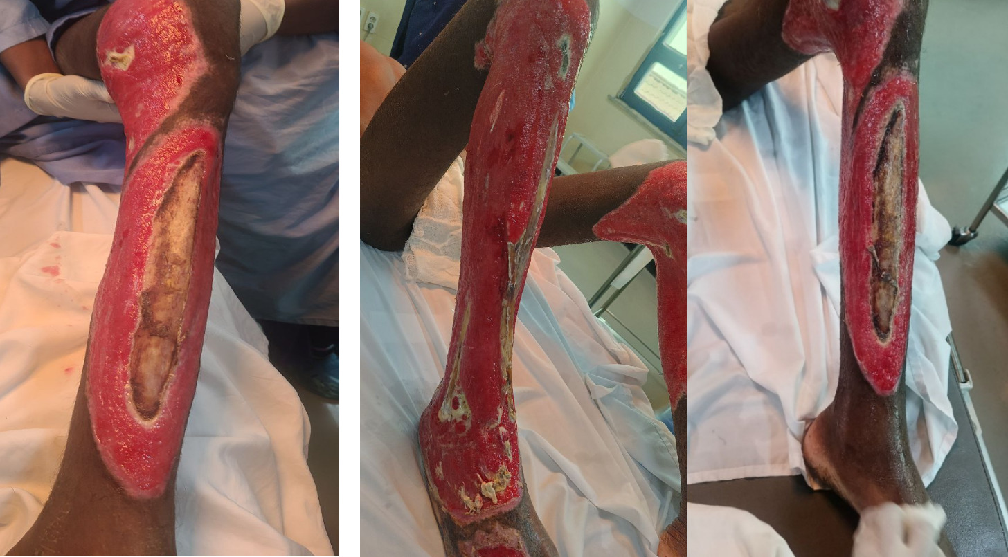
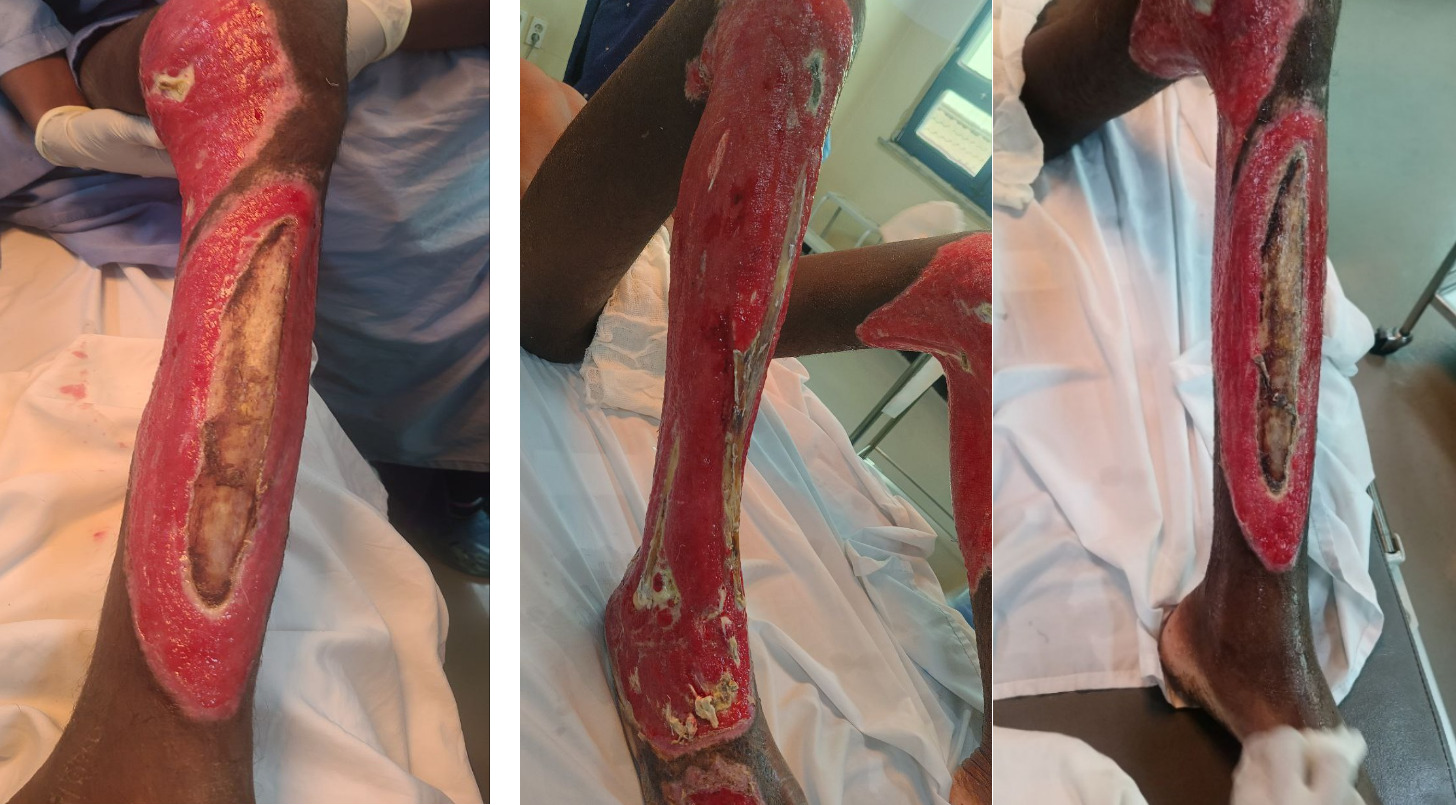
On admission, the patient appeared chronically ill-looking, cachectic, and in painful distress. He had extensive fourth-degree flame burns involving the entire lateral aspect of the right lower leg and the medial aspect of the left lower leg involved (Figure 1). The tibial bones were exposed bilaterally, the right fibula was exposed, tendons were exposed, soft tissue was missing, and there were areas of necrotic tissue and unhealthy granulation tissue (Figure 1).
Laboratory investigations demonstrated findings consistent with severe inflammation, anemia of critical illness, protein-calorie malnutrition, and hyper metabolic burn physiology.10–12
The patient was admitted in the burn and reconstructive surgery unit. Aggressive multidisciplinary management was initiated immediately to include the following:
-
High-calorie burn metabolic diet
-
Nutritional support guided by the Curreri formula
-
Propranolol therapy for persistent burn-related tachycardia
-
Multiple blood transfusions
-
Broad analgesia and perioperative pain control
-
Serial wound care and infection prophylaxis
Surgical Management
First operation: Initial radical debridement and cortical tibial trephination
The patient initially underwent extensive excisional debridement of bilateral lower extremity burn wounds under general anaesthesia. Intraoperative, large areas of necrotic skin, subcutaneous tissue, devitalized fascia, and nonviable soft tissue were identified over the lateral aspect of the right lower leg and medial aspect of the left lower leg. Bilateral exposure of the anterior tibial cortex and partial exposure of the right fibula were noted.
Given the extensive cortical exposure and absence of adequate vascularized wound bed, multiple cortical drill holes (cortical trephination technique) were created over both tibiae using a bone drill to stimulate medullary bleeding and promote granulation tissue formation over the exposed bone surface.7 Aggressive irrigation and wound bed preparation were performed, followed by application of moist burn dressings.
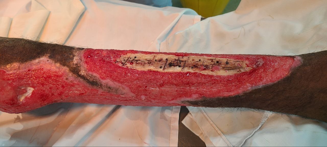
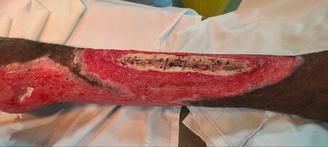
Second operation: Repeat debridement and additional cortical fenestration. (Figure 2)
A second staged procedure was performed due to persistent areas of nonviable tissue and exposed cortical bone. We performed a repeat excisional wound debridement, bilateral anterior tibial cortical drilling/fenestration, right fibular bone debridement, and wound bed preparation for future flap coverage.
Intraoperatively, early granulation tissue formation was observed arising from previously trephined cortical sites. Additional cortical fenestrations were made to further enhance vascular granulation over exposed tibial and fibular surfaces.
This staged approach was performed as part of limb salvage reconstruction prior to definitive soft tissue coverage.
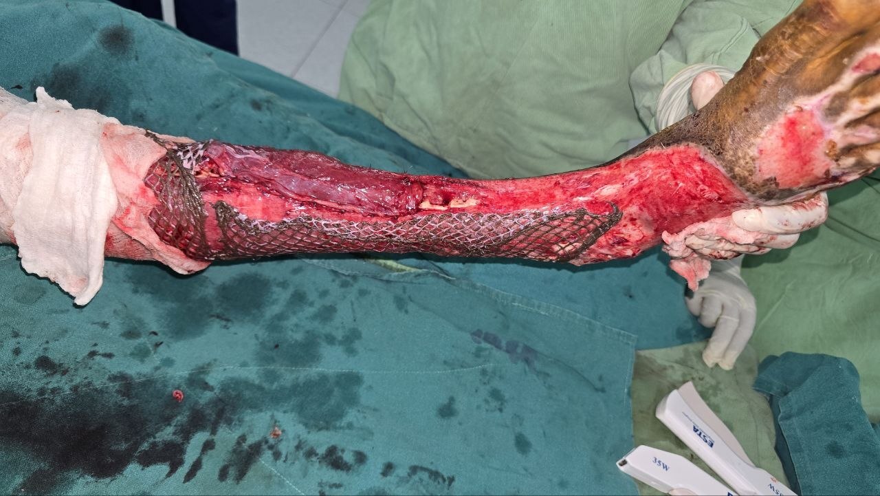
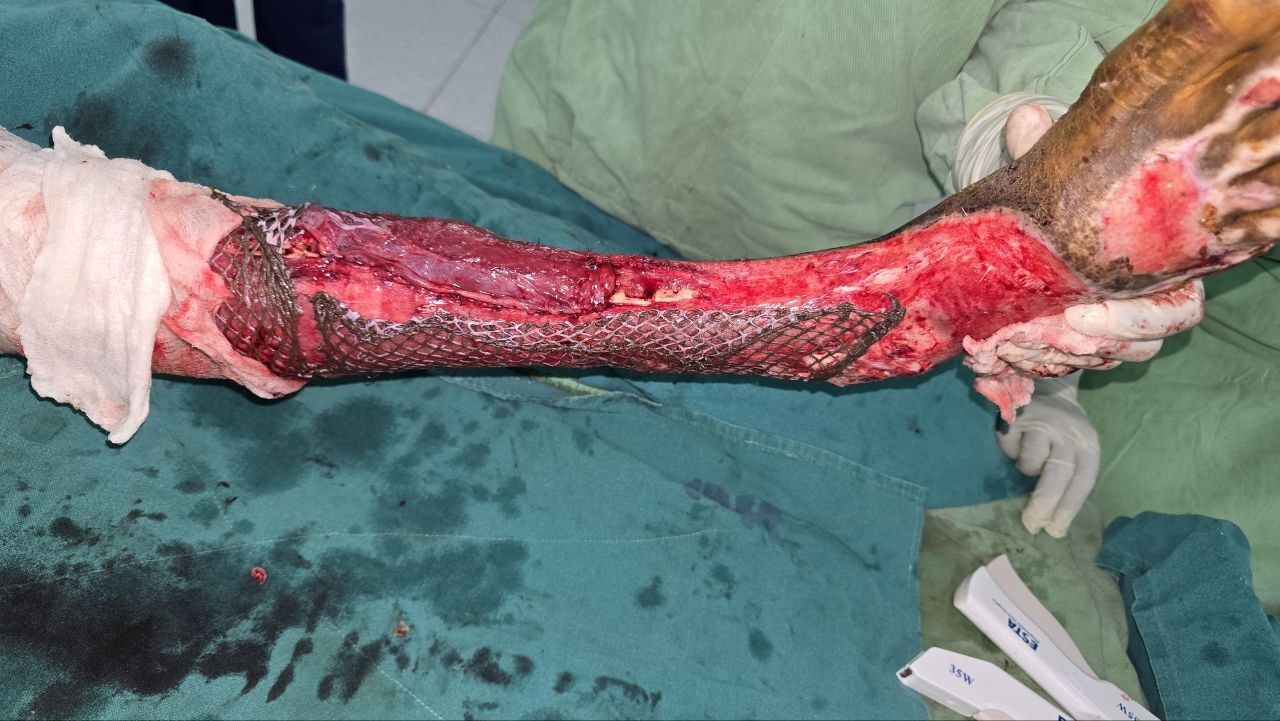
Third Operation: Right hemi soleus muscle flap reconstruction with meshed split-thickness skin grafting. (Figure 3)
Following successful granulation tissue development and infection control, definitive soft tissue reconstruction of the right lower extremity was undertaken. We performed excisional debridement and wound bed optimization, identification of the gastrocnemius-soleus complex, pedicled right hemisoleus muscle flap elevation, local muscle flap transposition for tibial coverage, and meshed split-thickness skin grafting (STSG). The medial hemisoleus muscle flap was mobilized based on its distal segmental vascular pedicles and rotated anteriorly to cover approximately 74% of the exposed tibial cortex. The flap was inset using interrupted absorbable sutures to achieve stable vascularized coverage of the exposed bone.
Subsequently, a meshed 1:4 split-thickness skin graft harvested from the ipsilateral thigh was applied over the muscle flap and remaining granulating wound bed.
This reconstructive strategy functioned similarly to a local rotational muscle flap procedure used for middle-third tibial reconstruction.13–16
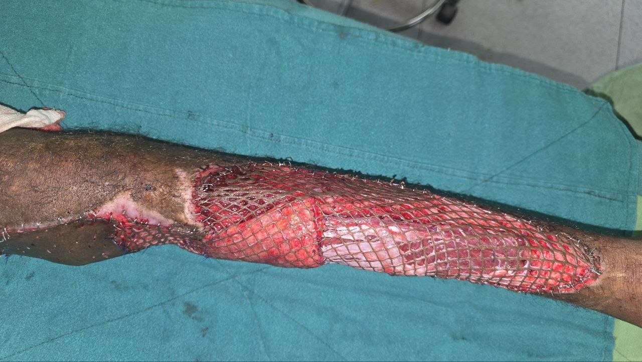
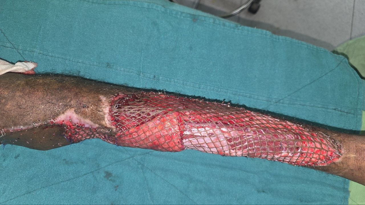
Fourth Operation: Left medial gastrocnemius–hemi soleus combined flap reconstruction with STSG. (Figure 4)
Definitive reconstruction of the left lower extremity was later performed. We performed sharp excisional debridement of unhealthy tissue, revision of previous hemisoleus muscle flap, medial gastrocnemius muscle flap advancement, combined gastrocnemius–soleus regional flap coverage, meshed STSG, popliteal fossa sheet grafting, and immobilization with dorsal plaster of Paris splint.
The medial gastrocnemius flap was mobilized proximally and advanced distally to augment tibial coverage. The hemisoleus flap was revised and inset together with the gastrocnemius flap to maximize vascularized soft tissue coverage over exposed bone. A 1:4 meshed split-thickness skin graft harvested from the ipsilateral thigh was then applied over the reconstructed wound. Additional sheet grafting was performed over the popliteal region to improve contour and minimize secondary contracture formation.
The wounds were dressed using Vaseline gauze and dry sterile gauze dressings, and a dorsal POP splint was applied for flap protection and immobilization.
Outcome and Follow-Up
Following completion of staged reconstructive procedures, the patient remained admitted under the burn and reconstructive surgery service for prolonged inpatient management, intensive wound monitoring, nutritional rehabilitation, pain control, infection surveillance, and physiotherapy.
During the postoperative period, the patient underwent daily wound assessment and dressing changes under strict aseptic precautions. Wound care consisted of Vaseline gauze application, dry sterile dressing reinforcement, graft inspection, flap viability monitoring, and limb immobilization using dorsal plaster of Paris (POP) splints to minimize shear stress over the reconstructed areas.
Serial postoperative evaluations demonstrated progressive improvement in both lower extremities. The hemisoleus and medial gastrocnemius muscle flaps remained viable with preserved vascularity and no evidence of flap necrosis, venous congestion, or ischemic compromise. The split-thickness skin grafts demonstrated satisfactory graft uptake over the majority of reconstructed wound surfaces with progressive epithelialization observed during subsequent dressing changes.
The previously exposed tibial and fibular cortices became adequately covered with vascularized tissue, and healthy granulation tissue continued to develop in residual small uncovered areas. No gross purulent discharge, progressive cellulitis, or signs of invasive soft tissue infection were observed following definitive reconstruction.
The patient’s systemic condition also improved gradually during hospitalization. Tachycardia decreased progressively following nutritional optimization, pain control, and propranolol therapy. Inflammatory status clinically improved with reduction in wound exudate and improvement in overall appearance of the wounds. Nutritional rehabilitation with high-calorie, high-protein burn diet resulted in improvement in appetite, general strength, and functional recovery.
Multiple blood transfusions administered during admission contributed to stabilization of symptomatic anemia. Serial monitoring showed clinical improvement in pallor, exercise tolerance, and overall physical endurance.
Pain management remained a major component of treatment because of repeated dressing changes, extensive tissue injury, and multiple staged surgical interventions. Multimodal analgesia was utilized throughout admission, allowing improved tolerance of wound care and early mobilization efforts.
As wound healing progressed, gradual physiotherapy and limb mobilization were initiated to reduce joint stiffness, muscle wasting, and contracture formation. Range-of-motion exercises for the knees and ankles were progressively introduced while protecting grafted and reconstructed areas.
The patient remained hospitalized for nearly two months because of the severity of injury, complexity of reconstruction, need for serial wound care, nutritional depletion, and prolonged recovery period.
By the time of discharge both lower limbs were successfully salvaged, muscle flaps remained viable, majority of skin grafts had successfully taken, tibial exposure had been adequately covered, no major postoperative complications requiring reoperation were identified, the patient was clinically stable, oral intake and nutritional status had improved, pain was adequately controlled with oral medications, and the patient was able to ambulate with assistance.
The patient was discharged with scheduled outpatient burn clinic follow-up, continued physiotherapy recommendations, nutritional supplementation advice, instructions for ongoing wound care and dressing changes, monitoring for possible late complications including contracture, graft breakdown, chronic osteomyelitis, hypertrophic scarring, and functional limitation
Long-term follow-up was planned to assess functional limb recovery, ambulation status, scar maturation, need for secondary reconstructive procedures, late contracture release if required, and psychosocial reintegration and quality of life improvement.
Despite delayed presentation, extensive bilateral fourth-degree burns, prolonged tissue exposure, and limited initial burn care resources, the patient achieved favorable limb salvage and wound healing outcomes through aggressive multidisciplinary burn management and staged reconstructive surgery.
Discussion
Fourth-degree burns represent one of the most devastating forms of thermal injury and are characterized by destruction extending beyond the skin into subcutaneous tissue, fascia, muscle, tendon, and bone.1,4 These injuries are uncommon compared with superficial and partial-thickness burns but carry substantially higher rates of morbidity, prolonged hospitalization, infection, disability, and limb loss.1,2 The present case illustrates the complexity of managing extensive bilateral lower extremity fourth-degree flame burns with exposed tibia and fibula in a resource-limited setting and highlights the role of staged limb salvage reconstruction using cortical bone drilling, regional muscle flaps, and split-thickness skin grafting.
Flame burns remain a major cause of severe burn injury worldwide, particularly in rural and low-income populations where open-fire exposure is common.2,3 Young adult males are disproportionately affected because of occupational and environmental exposure risks.3 In the present case, the patient sustained injury while camping and tending camels in a rural environment, illustrating the continued vulnerability of pastoral communities to severe thermal trauma.
One of the major factors contributing to the severity of this patient’s condition was delayed definitive management. The patient remained at the referring institution for 23 days before transfer to a specialized center, during which wound care was limited due to lack of adequate sedation and reconstructive resources. Delayed excision and inadequate wound coverage are known to increase the risk of progressive necrosis, invasive infection, desiccation of exposed structures, systemic inflammatory response, and chronic osteomyelitis.5,7,8 Early referral to specialized burn centers has consistently been associated with improved outcomes, lower infection rates, shorter hospital stay, and reduced need for amputation.1,4
The lower extremity, particularly the anterior tibial region, presents a unique reconstructive challenge because of minimal soft tissue coverage over bone.7 Once the periosteum is destroyed and cortical bone becomes exposed, spontaneous granulation tissue formation is severely impaired due to poor vascularity of the cortical surface.7 In such situations, conventional skin grafting alone is usually unsuccessful because graft survival depends on a well-vascularized recipient bed.6
To address this problem, cortical drilling (also referred to as cortical fenestration or trephination) was performed in this patient. This technique involves creating multiple perforations through the outer cortex to expose the vascular medullary cavity and stimulate granulation tissue formation.7 The procedure is particularly valuable in resource-limited settings where advanced reconstructive modalities such as free tissue transfer or dermal substitutes may not be readily available. In the present case, progressive granulation tissue formation following serial cortical drilling facilitated subsequent flap coverage and graft take.
Serial debridement also played a critical role in management. Burn wound excision removes necrotic tissue, decreases bacterial burden, reduces inflammatory mediators, and prepares the wound bed for reconstruction.1,18 In extensive fourth-degree burns, repeated debridement is frequently necessary because tissue viability evolves over time and deeper necrosis may become demarcated gradually.18 In our patient, staged debridement allowed progressive wound bed optimization prior to definitive reconstruction.
Definitive soft tissue coverage was achieved using pedicled hemisoleus and medial gastrocnemius muscle flaps combined with split-thickness skin grafting. Muscle flaps remain among the most reliable reconstructive options for exposed tibial bone because of their robust vascularity, ability to obliterate dead space, resistance to infection, and promotion of wound healing.6,13–16
The hemisoleus flap is particularly useful for middle-third tibial defects due to its segmental blood supply and adequate arc of rotation.13–16 The soleus muscle can be divided longitudinally while preserving partial muscle function, making the hemisoleus flap especially advantageous for moderate-sized defects involving the middle leg.15 In this patient, the hemisoleus flap successfully covered approximately 74% of the exposed anterior tibial surface on the right lower extremity, providing durable vascularized coverage and facilitating successful graft take.
Similarly, the medial gastrocnemius flap remains one of the most dependable local flaps for proximal and upper-middle tibial defects.14 Its rich vascular supply from the sural branches of the popliteal artery provides reliable tissue perfusion even in previously traumatized extremities.8,14 In this case, combination use of the hemisoleus and medial gastrocnemius flaps allowed expanded coverage of extensive bilateral defects while avoiding more technically demanding microsurgical free flap procedures.
Alternative reconstructive options for lower extremity burn defects include reverse sural fasciocutaneous flaps, perforator flaps, cross-leg flaps, and free tissue transfer.8,17 Reverse sural flaps are commonly utilized for distal tibial, ankle, and heel defects because of their relatively simple technique and preservation of major vascular structures.17 However, their reliability decreases in extensive burn injuries because of compromised perforator circulation, surrounding tissue damage, and increased risk of venous congestion.17 In the current patient, the extent and location of tissue loss favored regional muscle flap reconstruction over fasciocutaneous alternatives.
Free flap reconstruction is considered the gold standard for extensive lower extremity soft tissue loss in many tertiary centers because it provides large volumes of vascularized tissue.16 However, microsurgical reconstruction requires advanced expertise, prolonged operative time, specialized postoperative monitoring, and substantial healthcare resources.16 These factors may limit its feasibility in resource-constrained settings. The successful use of regional pedicled muscle flaps in this patient therefore demonstrates the effectiveness of practical reconstructive alternatives in low-resource environments.
Another major aspect of management in this patient was metabolic and nutritional support. Severe burns induce a profound hypermetabolic state characterized by catecholamine excess, persistent tachycardia, insulin resistance, protein catabolism, muscle wasting, immune dysfunction, and increased resting energy expenditure.10,11 Without aggressive nutritional support, patients rapidly develop protein-calorie malnutrition, impaired wound healing, and increased susceptibility to infection.12
Our patient demonstrated clinical and biochemical evidence of severe metabolic stress, including tachycardia, hypoalbuminemia, low total protein levels, anemia, and elevated inflammatory markers. Nutritional rehabilitation using a high-calorie burn diet guided by the Curreri formula was therefore initiated immediately. The Curreri formula remains a commonly used approach for estimating caloric requirements in burn patients, particularly in settings where indirect calorimetry is unavailable.1 Propranolol therapy was also administered to attenuate the hypermetabolic response. Beta-blockade in severe burn injury has been shown to reduce resting energy expenditure, cardiac stress, muscle protein breakdown, and peripheral lipolysis.10,11 Herndon et al. demonstrated that propranolol significantly reverses burn-associated catabolism and improves physiologic recovery.11 In the present case, gradual improvement in tachycardia and overall clinical status was observed following combined nutritional and pharmacologic metabolic management.
Infection prevention represented another essential component of care. Burn wounds are highly susceptible to bacterial colonization because of loss of the protective skin barrier, impaired immunity, and prolonged hospitalization.18 Extensive lower extremity wounds with exposed bone further increase the risk of osteomyelitis and systemic sepsis.7,8 Although wound cultures were not available in this report, serial debridement, dressing care, nutritional support, and timely flap coverage likely contributed to prevention of overwhelming infection and successful limb salvage.
The prolonged hospitalization period of nearly two months was expected given the severity of tissue destruction, need for multiple surgeries, daily wound care, nutritional rehabilitation, and rehabilitation therapy. Long-term complications following severe lower extremity burns may include hypertrophic scarring, chronic pain, joint contractures, gait impairment, psychological distress, and recurrent ulceration.1,8 Continuous follow-up and rehabilitation are therefore essential for optimization of functional outcomes.
This case is particularly important because it demonstrates that successful limb salvage is achievable even in severely advanced fourth-degree burns within resource-limited healthcare systems. Despite delayed referral, prolonged tissue exposure, extensive bilateral defects, and limited initial burn care, multidisciplinary management using staged debridement, cortical drilling, regional muscle flaps, and split-thickness skin grafting resulted in preservation of both lower limbs.
The case also highlights several important lessons:
-
Early transfer to specialized burn centers is critical
-
Serial debridement and infection control are essential
-
Cortical bone drilling remains a valuable technique for exposed tibial wounds
-
Regional muscle flaps provide reliable reconstruction in resource-limited settings
-
Nutritional and metabolic support significantly influence wound healing outcomes
-
Multidisciplinary burn care can achieve limb salvage even in highly complex injuries
Overall, this report contributes to the limited literature describing successful reconstruction of bilateral fourth-degree lower extremity burns with exposed tibia in low-resource settings and may help guide burn surgeons facing similar reconstructive challenges.
Ethics approval and consent to participate
Ethical approval for publication of this case report was obtained from the Myungsung Medical College Institutional Review Board (IRB)/Ethics Committee, Addis Ababa, Ethiopia. All procedures were performed in accordance with the ethical standards of the institution and the principles of the Declaration of Helsinki.
Consent for publication
Written informed consent was obtained from the patient for publication of this case report and the accompanying clinical images. A copy of the written consent is available for review by the Editor-in-Chief of this journal upon reasonable request.
Competing interests
The authors declare that they have no competing interests.
Funding
The authors received no financial support, grants, or funding for the preparation, authorship, or publication of this case report.
Acknowledgements
The authors sincerely thank the Burn and Reconstructive Surgery team, nursing staff, anaesthesiology team, physiotherapists, and all healthcare professionals involved in the multidisciplinary care of the patient. The authors are also grateful to the patient for providing consent to share his clinical course for educational and scientific purposes.