Background
Panophthalmitis is characterized by the extension of intraocular infection into the sclera, Tenon’s capsule, and adjacent orbital soft tissues.1 Panophthalmitis presents with signs of orbital involvement, such as proptosis, chemosis, and ophthalmoplegia.1 As the infection involves vascularized tissues, the treatment of panophthalmitis is aided by intravenous antibiotics.2 Systemic treatment is mandatory to treat the primary source due to the risk of sepsis and other systemic infectious complications.2 Furthermore, repeated intravitreal injections of broad-spectrum antibiotics, such as vancomycin and ceftazidime, are often required to control the intraocular component. In severe cases, orbital inflammation can lead to the development of cavernous sinus thrombosis (CST).3 This progression is facilitated by the region’s venous anatomy; specifically, the valveless nature of the superior and inferior ophthalmic veins, coupled with elevated intraocular pressure, promote the drainage of infectious venous blood into the dural sinuses.3,4 When panophthalmitis evolves to include CST, immediate enucleation is considered the standard of care to ensure complete eradication of the infectious source and devitalized tissues.1,5 Even with surgical debridement and aggressive antibiotic treatment, CST carries a mortality rate of up to 30%, and survivors may have permanent sequelae, such as cranial neuropathies.6
The decision to enucleate poses an emotional and psychological challenge for patients and their families to accept. However, clinicians often emphasize that in the setting of panophthalmitis and CST, globe retention may constitute a direct threat to the patient’s life. Successful management of these concurrent conditions with only medical management and globe-sparing surgery is exceedingly rare, given the high rates of morbidity and mortality. This report presents a unique case of panophthalmitis with associated CST where globe preservation was successfully achieved at the patient’s insistence.
Case Presentation
A 69-year-old woman with a history of hypothyroidism presented to the emergency department (ED) with worsening ocular pain, edema, erythema, purulent drainage, and sudden loss of vision in the left eye. Three weeks prior to presentation, the patient was evaluated by an outside retinal specialist for floaters, tearing, and blurry vision. At that initial visit, her best corrected visual acuity was measured at 20/70 in the right eye (OD) and 20/100 in the left eye (OS), and she was found to have a severe increase in intraocular pressure (IOP). An anterior chamber tap was performed to reduce the elevated IOP. Two weeks later, the patient developed fulminant endophthalmitis, prompting her ED visit.
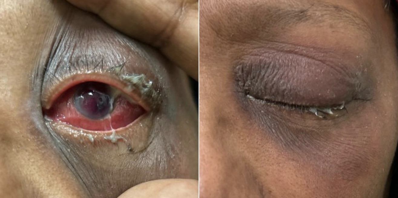
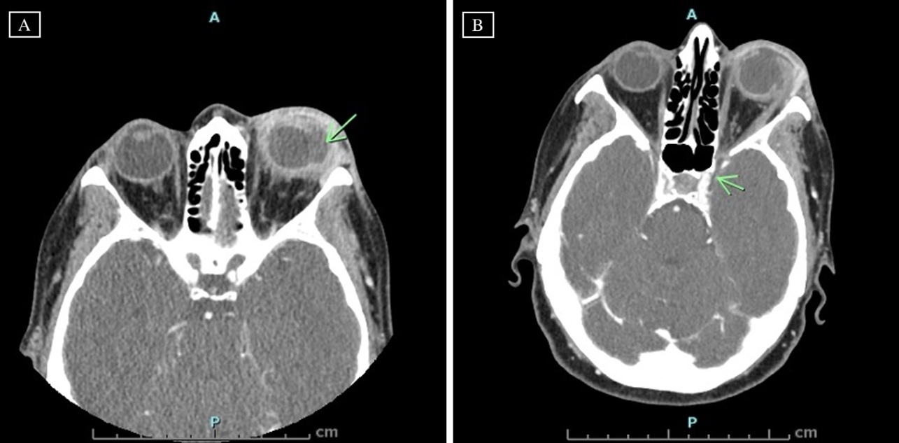
Upon evaluation in the ED (day 1), best corrected visual acuity in the left eye was determined to be strictly no light perception (NLP). She denied any history of ocular trauma or prior eye surgeries. The patient expressed no concerns regarding the right eye. Slit-lamp examination of the left eye was notable for 2+ injection, superotemporal subconjunctival purulent discharge through what appeared to be ruptured sclera, and a layered hyphema of the anterior chamber; consequently, neither the lens nor the posterior segment could be visualized (Figure 1). Orbital computed tomography (CT) scan demonstrated severe left-sided orbital infection and intraocular inflammation. CT venography of the head revealed cavernous sinus thrombosis (Figure 2). The patient was diagnosed with panophthalmitis complicated by cavernous sinus thrombosis. Following admission for intensive IV antibiotic management, conjunctival cultures from the area of purulent drainage identified Streptococcus intermedius susceptible to ceftriaxone.
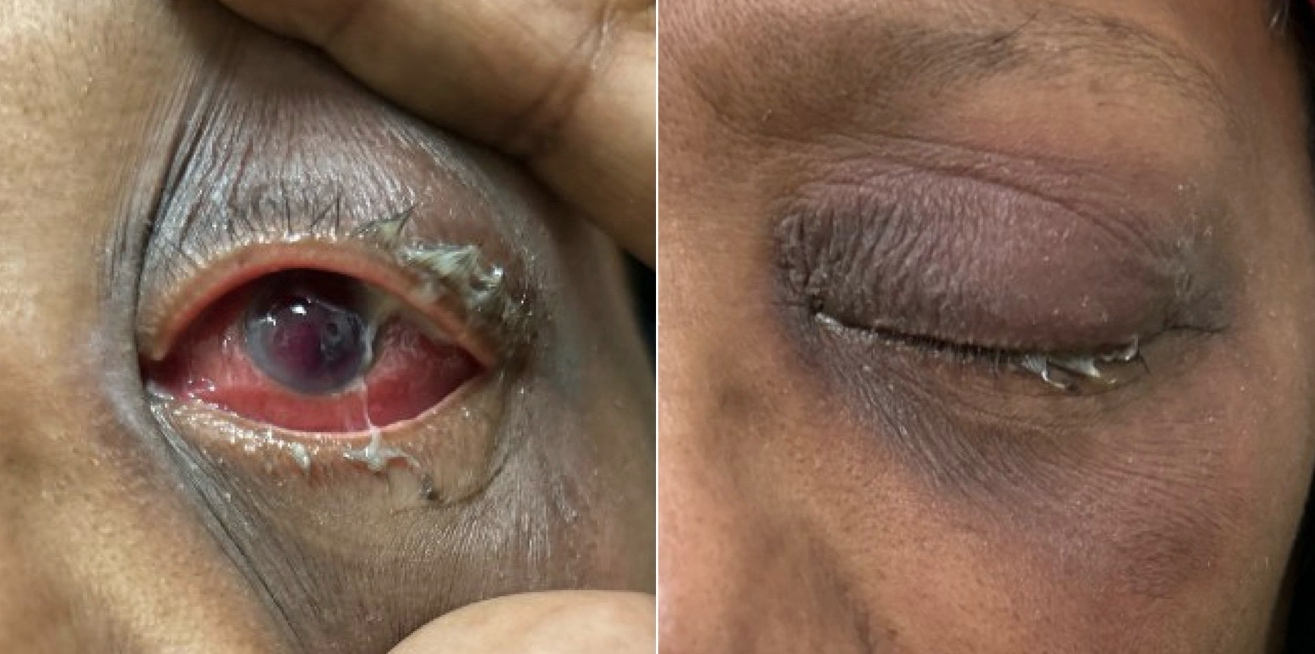
_at_presentation__ct_of_the_orbits_with_iv_contrast_demonstrat.jpeg)
Intravenous ceftriaxone (2 g every 12 hours) was initiated for a planned six-week course and a peripherally inserted central catheter was placed. Adjunctive topical therapy for the left eye included moxifloxacin drops every three hours and mupirocin 2% ointment twice daily. The patient was counseled regarding the critical risks associated with a non-visual eye in the setting of orbital infection and cavernous sinus involvement. The care team emphasized that further local or systemic spread could be life-threatening and recommended enucleation as part of definitive treatment; however, the patient declined the procedure, stating a strong desire to preserve the eye. Over the remainder of her hospitalization, the patient demonstrated steady clinical improvement on the antimicrobial regimen; her presenting ocular pain resolved completely, accompanied by a marked decrease in both periorbital swelling and purulent drainage. At discharge, the patient was provided with strict return precautions and instructed to seek immediate emergency care for fever, increased swelling, ocular pain, or any clinical deterioration.
The patient continued close follow-up with both the ophthalmology and infectious disease teams and underwent follow up orbital CT scans to ascertain any worsening of infectious spread. The patient remained firm in the decision to decline enucleation, expressing a strong preference for globe preservation despite repeated counseling on the risks. Over the subsequent weeks, serial slit-lamp examinations demonstrated overall clinical stability, characterized by a gradual improvement in periorbital edema and anterior segment inflammation, though the posterior segment remained obscured. Given the continued clinical improvement with medical management, the current regimen of intravenous ceftriaxone and topical moxifloxacin was continued.
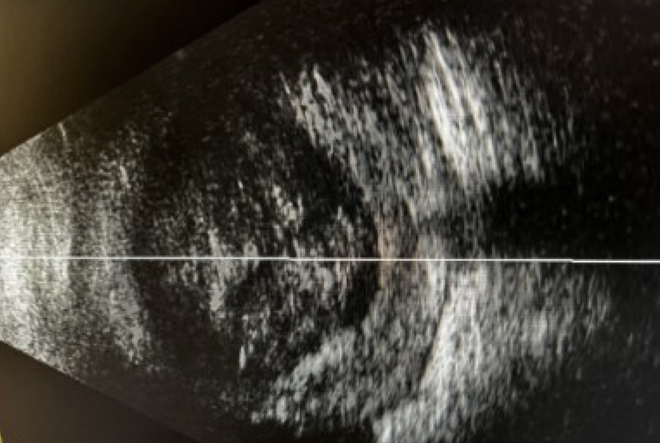
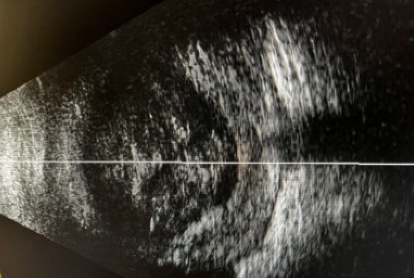
On day 12, a retinal specialist performed a vitrectomy and anterior chamber washout of the left eye. Intravitreal vancomycin, voriconazole, and ceftazidime were administered at the conclusion of the procedure. Vitreous cultures yielded no growth, and the systemic ceftriaxone and topical moxifloxacin regimen was maintained. Postoperatively, the patient reported a significant reduction in pain compared to the initial presentation. On slit-lamp examination, necrotic sclera was noted superotemporally. A B-scan ultrasound of the left eye revealed diffuse vitreous opacities and thickened sclera (Figure 3). On day 23, repeat CT of the orbit showed interval improvement in inflammatory changes noted around the globe and sclera, unremarkable retrobulbar and intraconal fat, no change in cavernous sinus, no enlargement of superior ophthalmic veins, and no intracranial extension. Per recommendations by the infectious disease team, the intravenous ceftriaxone dosage was reduced from 2 g every 12 hours to every 24 hours, while daily topical moxifloxacin drops were continued.
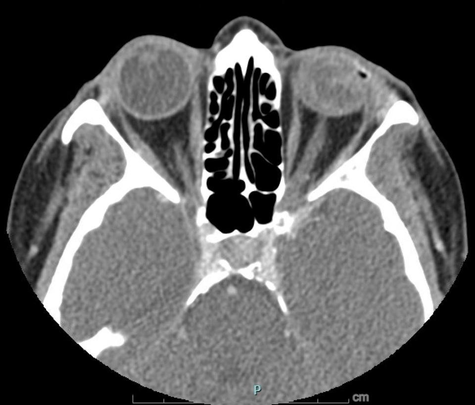
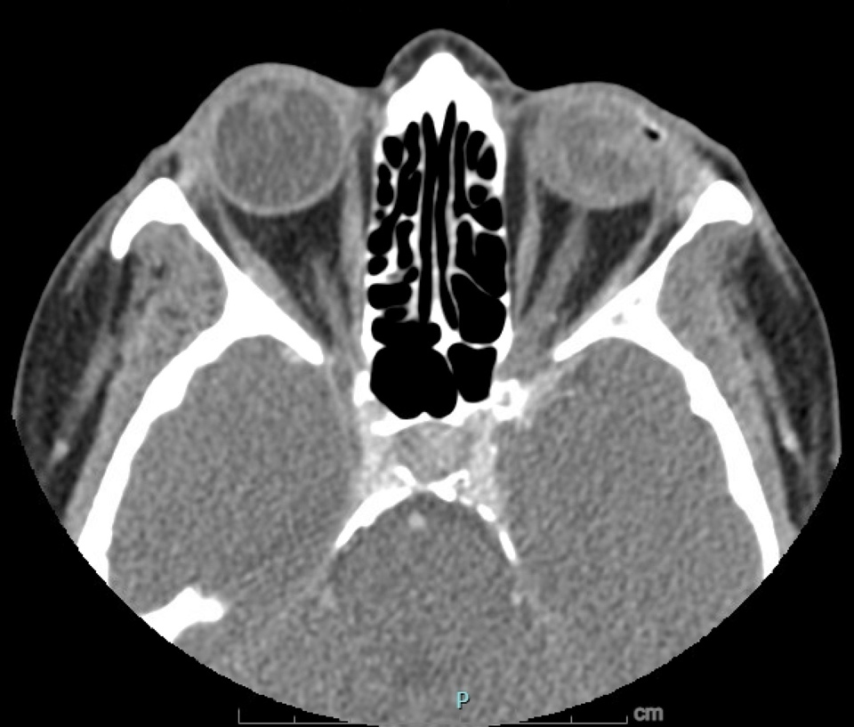
On day 66, CT of the face revealed a stable, heterogeneously dense, and shrunken left globe. The left cavernous sinus showed improvement in the filling defect, suggestive of evolving sequelae from the thrombosis. Clinically, the left eye showed signs of phthisis bulbi and visual acuity of no light perception. Twice daily atropine drops were prescribed, and topical moxifloxacin was increased from a once daily dose to four times. On post-presentation day 121, the patient denied ocular pain, headaches, focal weakness, or orbital tenderness. There was no evidence of recurrent or persistent infection (Figure 4). Slit-lamp examination of the left eye was notable for enophthalmos, ptosis, and a white and quiet conjunctiva with a localized superotemporal scar. The cornea demonstrated reduced sensation, peripheral neovascularization, and scattered epithelial defects. The anterior chamber remained deep and quiet with evidence of scattered iris atrophy; however, there was no view of the lens or posterior segment due to a pupillary membrane. The examination was consistent with a pre-phthisical globe with no remaining visual potential.
She expressed a desire for improved cosmesis, and management options, such as a scleral shell, enucleation with a dermis-fat graft, or continued observation, were discussed in detail. The patient remains under the care of the ophthalmology service for long-term management. This case highlights the importance of persistent treatment and multidisciplinary intervention in preventing life-threatening intracranial extension of panophthalmitis, even when the visual prognosis remains poor.
Discussion
Endophthalmitis is characterized as inflammation of the intraocular structures resulting from infectious colonization.7 Endophthalmitis is a serious and vision-threatening condition, and as such, prompt intervention is crucial.7 Endophthalmitis can be classified as exogenous or endogenous based on the mode of transmission of the infectious source. Exogenous endophthalmitis occurs via direct inoculation of the organisms through surgery, trauma, or adjacent tissues. Contrastingly, endogenous endophthalmitis occurs when infectious organisms spread via a hematogenous route to the eye from a distant location.7 Treatment of endophthalmitis often begins with a vitreous sample for culture with concomitant intravitreal injection of antibiotics.8 Systemic antibiotics may be used as the primary treatment or alongside local therapy. However, due to the blood-retinal barrier limiting the intraocular penetration of some agents, selecting systemic agents with high ocular bioavailability is critical. In more aggressive cases, a pars plana vitrectomy, which reduces the infectious load and provides a diagnostic sample, is recommended.8
While endophthalmitis is confined to the intraocular cavities, panophthalmitis extends beyond the structural integrity of the globe, essentially converting the eye into a reservoir of infection. Complications of panophthalmitis are diverse and include meningitis, brain abscesses, subdural empyema, pituitary apoplexy, septic pulmonary emboli, sympathetic ophthalmia, orbital apex syndrome, and more.2,3 Interestingly, these complications were not experienced by this patient throughout the treatment course.
Due to the clinical scenario of panophthalmitis and its defiance to medical treatment alone, the consensus in peer-reviewed literature is that management of panophthalmitis requires antibiotics and surgical removal of the globe for the most complete infectious debridement.5 As panophthalmitis involves scleral infection, evisceration, which leaves the sclera behind, is inadequate. Therefore, enucleation is often the definitive surgical approach to ensure complete eradication of the infectious source and associated devitalized tissue.5 Antibiotics alone are rarely sufficient to penetrate a necrotic, pus-filled globe or septic thrombi effectively enough to remove the bacterial burden. Due to the underlying risk of sepsis, panophthalmitis has a high risk of mortality, which emphasizes the need for prompt recognition and treatment. The goal is to remove the bacterial burden before patients evolve from panophthalmitis to further spread of infection, such as CST.
The presentation of panophthalmitis coupled with CST is a complex and life-threatening infectious condition. The structure of the cavernous sinus makes it favorable for bacterial colonization and thrombus formation, increasing the patient’s risk of mortality and morbidity.4 Formation of a thrombus within the sinus can occur due to the high concentration of inflammatory cytokines, which create a local hypercoagulable environment.3 CST often presents with chemosis, periorbital edema, pain, and vision loss. CST is diagnosed through neuroimaging, and treatment requires high-dose intravenous antibiotics, anticoagulation, and surgical removal of the source.3 With a diagnosis of CST, mortality jumps to 30% for treated cases and 100% if left untreated.4 Additionally, approximately 50% of survivors of cavernous sinus thrombosis are left with other severe and chronic complications,4 including cranial nerve palsies, persistent diplopia, and facial numbness, which were not reported in this patient. However, in alignment with current statistics showing 22% of survivors having significant visual impairment or blindness, this patient’s visual acuity remained at no light perception.9 The evolution of endophthalmitis to panophthalmitis and ultimately CST illustrates a dangerous trajectory facilitated by the breakdown of ocular barriers and the unique valveless venous anatomy of the orbit. The literature suggests that enucleation is important not only for pain relief but as a life-saving intervention.
Conclusions
This case demonstrates the successful resolution of panophthalmitis and concomitant CST through medical management and globe-sparing surgery. The outcome is particularly noteworthy; a vitrectomy and aggressive antimicrobial therapy not only eradicated the primary infection and prevented recurrence but also achieved globe preservation. Despite the lack of surgical enucleation, traditionally considered the standard for source control in such advanced cases, the patient remained free of further systemic or orbital complications. While this case represents a unique instance in which antibiotics and a vitrectomy alone were sufficient to treat the patient’s diagnosis of panophthalmitis and CST, it is important to note that it should not influence standard of care guidelines. Ultimately, this case serves as a remarkable clinical outlier of successful medical management due to strong patient preference against surgical debridement.
Conflicts of Interest
The authors declare that they have no conflicts of interest.
Funding
The authors received no financial support for the research, authorship, and/or publication of this article.
Informed Consent
Written informed consent was obtained from the patient for publication of this case report and any accompanying images. All information has been de-identified to protect the patient’s privacy.