Introduction
A urachus is a structure derived from the allantois, formed by the hindgut, which establishes a connection between the bladder and umbilicus during fetal development. This connection typically disappears around the end of the first trimester.1,2 It is typically composed of transitional epithelium; however, approximately 30% have columnar epithelium as the deepest layer. Surrounding the transitional or columnar epithelium is a layer of submucosal connective tissue, followed by a final layer of muscle.3 If the urachus fails to close properly, it can lead to potential complications throughout life, including a patent urachus, urachal cysts, and vesicourachal diverticulum, among others.4 The incidence of all urachal anomalies is 1/5000.5
There is also a risk of cancer if the urachus does not close completely, leaving behind some form of urachal remnant. Urachal cancer is typically an adenocarcinoma.6 It has a 5-year cancer-specific survival rate of approximately 35%.7
Urachal anomalies can present with various symptoms including umbilical drainage, abdominal pain, an erythematous umbilicus, and recurrent urinary tract infections.2,4 The incidence of a urachal anomaly that also communicates with the sigmoid colon, as seen in this case, is even rarer, with very few cases reported in the literature.8 The pathology described here involves a fistulous connection between the bladder, umbilicus, and the sigmoid colon, complicated by diverticulosis. The following discussion includes the presentation, imaging, surgical intervention, and outcomes associated with this case of a urachal-sigmoid fistula.
Case Description
A 44-year-old male with a past medical history of hypertension and diverticulosis complicated by diverticulitis presented with two weeks of umbilical drainage, erythema, and pain surrounding the umbilicus. The umbilical drainage was at times bloody, purulent, feculent, and contained urine and air. On admission, he reported a temperature as high as 101.5°F, along with night sweats and dysuria. His vital signs were as follows: blood pressure 129/101 mmHg, pulse 67 beats per minute, temperature 98.1°F, respirations of 16 breaths per minute, and oxygen saturation (SpO2) of 96%. His body mass index (BMI) was 34.2. The only laboratory abnormality noted was a slightly elevated lactic acid level of 2.3 mmol/L, and there was no evidence of leukocytosis. A urinalysis revealed proteinuria, with no other abnormalities detected.
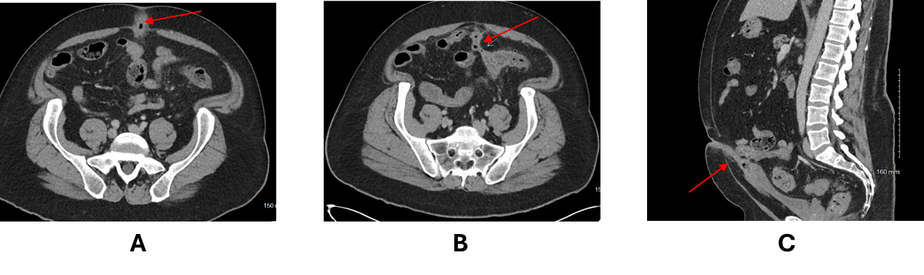
A CT scan revealed an elongated bladder in the midline, along with a fistulizing connection between the umbilicus and the sigmoid colon. A diagnosis of a urachal-sigmoid fistula was established (Figures 1). The patient was started on ciprofloxacin, metronidazole, and neomycin, which provided immediate relief. The patient was bowel prepped for resection, during which he experienced copious liquid discharge from the umbilicus. The procedure began as a laparoscopic resection but was converted to open due to the size and extent of the urachus. The urachus was carefully dissected and removed down to the base of the bladder, followed by sigmoid resection with primary anastomosis. The patient was discharged on postoperative day 5 and was doing well at his outpatient follow-up.
_and_sagittal_(c)_ct_images_depicting_the_urachal-sigmoid_fistula_(arro.png)
Discussion
The urachus is used by a developing fetus as a connection between the bladder and umbilicus but should disappear before birth. Occasionally, when this does not occur, a persistent connection between these two structures may lead to complications for patients. This report presents a case of a urachal-sigmoid fistula associated with umbilical drainage as one such complication.
This is a rare occurrence, with few other cases reported in the literature. When compared to other types of diverticular and genitourinary fistulas, the incidence rates for colovesicular fistulas range from 2% to 23% in patients with concurrent diverticulosis.8
Most patients who have experienced this pathology exhibited symptoms similar to those of our patient, including drainage, abdominal pain, and an erythematous umbilicus. A literature review of other similar cases revealed that all patients with urachal-sigmoid fistulas were treated in a manner consistent with the case described here, involving the resection of the affected area of the sigmoid colon and the removal of the urachal remnant. The primary difference noted was whether an antibiotic course was administered prior to resection due to infection at the site.9–13
Another point of discussion is the type of resection required of the urachus. One study found that among patients identified with a urachal anomaly, 51% had urachal cancers.14 Additionally, when patients present with these malignancies, they are often advanced.15 Due to the characteristics associated with the incidence and prognosis of urachal cancers, it is crucial to ensure that they are completely resected down to the base of the bladder to ensure that the urachus has been fully removed to try to avoid potential complications.
Conclusions
This case describes the rare presentation, imaging findings, and treatment of a urachal-sigmoid fistula in adults. Given the limited number of documented cases in the literature, it is crucial to have case reports available to assist in the future diagnosis and treatment of patients with this condition.