

Introduction
Spigelian hernia is a rare type of anterior abdominal wall hernia accounting for approximately 0.12 to 2% of cases.1 Hernia neck is located lateral to semilunar line which extends from the tip of the nineth costal cartilage to the pubic spine.2 It is more commonly found infra-umbilically in a region known as the spigelian belt.2,3 Clinical presentation of a spigelian hernia can vary from asymptomatic cases to vague abdominal pain which, depends on the hernia’s location and the sac content, to an abdominal lump or features intestinal obstruction or strangulation.1,4 Even though a diagnosis can be made in clinical grounds alone, radiological adjuncts like ultrasound scan (USS) and CT are necessary for unusual cases.1,5,6 Treatment of a spigelian hernia is a surgical repair with open or laparoscopic modalities.1,4
Strangulation of a hernia occurs when blood supply through a narrow neck in compromised to the herniated content and can lead to catastrophic events like bowel gangrene, sepsis and potentially death.7 Classically strangulated hernias are painful due to ischemia and inflammation. However, we report an unusual presentation of a strangulated spigelian hernia as a painless abdominal lump.
Case Presentation
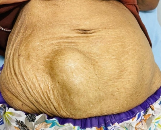
A 65-year-old previously well woman presented with an acute onset painless lump in the right lower abdomen for two days (Figure 1). This patient had no history of a trauma to the abdomen or had symptoms of intestinal obstruction.
On examination she was afebrile, and her haemodynamic parameters were within normal range. There was a non-tender compressible but non reducible lump in the right lower quadrant just lateral to the midline. Her blood investigation parameters including serum lactate were within normal range and USS revealed gas-filled content within the lump.
Immediate exploration through a transverse incision over the lump revealed a spigelian hernia with viable small bowel with a mildly cyanotic segment indicating early strangulation (Figure 2). The content was reduced, and the narrow neck of the hernia was repaired anatomically. Postoperative period was uncomplicated and she was discharged on the post operative day two. She returned for follow-up two weeks later with complete recovery.

Discussion
Diagnosis of spigelian hernia is straightforward when the classical clinical features are present. Strangulation can often be diagnosed or suspected on clinical grounds. However, diagnostic challenges can arise in atypical cases. Even though USS is operator dependant, it is reliable in most cases, whereas the CT remains the definitive modality.5,6 Both USS and CT can suggest strangulation by demonstrating reduced vascularity. Surgical repair is recommended for a diagnosed spigelian hernia to prevent future complications. Minimally invasive approaches such as transperitoneal or preperitoneal laparoscopic repair have gained popularity over conventional open surgery due to reduced post operative pain and quicker recovery.1,4,8 Although mesh repair is described to be safe in strangulated abdominal wall hernias, its role in spigelian hernia remains less well defined.9–11
In this case, diagnosis was supplemented with USS as a sudden appearance of a lump was an unusual feature of a hernia. Strangulation was neither suspected clinically as patient was completely pain free nor suggested at the USS. CT scan was not performed as USS confirmed the diagnosis. Immediate exploration was decided to prevent potential complications, and open surgery was performed as the available expertise and resources at that time. Since strangulation was present, anatomical repair was preferred to avoid potential mesh related complications. The absence of pain in this case could be attributed to early stage of strangulation or the higher pain threshold observed in elderly patients.12
Conclusions
Strangulated Spigelian hernia can present as a painless abdominal lump, highlighting the need for a high index of suspicion. Timely intervention is crucial to prevent catastrophic complications.
Acknowledgments
The subject gave informed consent, and patient anonymity was preserved. Authors wish to acknowledge the other medical staff who took care of the patient.